Email: info@neweastbio.com   |  Call: 6109452007

DAP3 Rabbit pAb

Anti-DAP3 Rabbit Polyclonal Antibody
5/5

$349.00

Cat.#:  S216471

   Size:   100 μl

In Stock

          Product Description          
Cat.#:   S216471
Product Name:   Anti-DAP3 Rabbit Polyclonal Antibody
Synonyms:   DAP-3; S29mt; MRPS29; MRP-S29; bMRP-10
UNIPROT ID:   P51398 (Gene Accession – BC107488 )
Background:    Mammalian mitochondrial ribosomal proteins are encoded by nuclear genes and help in protein synthesis within the mitochondrion. Mitochondrial ribosomes (mitoribosomes) consist of a small 28S subunit and a large 39S subunit. They have an estimated 75% protein to rRNA composition compared to prokaryotic ribosomes, where this ratio is reversed. Another difference between mammalian mitoribosomes and prokaryotic ribosomes is that the latter contain a 5S rRNA. Among different species, the proteins comprising the mitoribosome differ greatly in sequence, and sometimes in biochemical properties, which prevents easy recognition by sequence homology. This gene encodes a 28S subunit protein that also participates in apoptotic pathways which are initiated by tumor necrosis factor-alpha, Fas ligand, and gamma interferon. This protein potentially binds ATP/GTP and might be a functional partner of the mitoribosomal protein S27. Multiple alternatively spliced transcript variants encoding distinct isoforms have been found for this gene. Pseudogenes corresponding to this gene are found on chromosomes 1q and 2q.
Immunogen:    Fusion protein of human DAP3
Applications:    ELISA, WB, IHC
Recommended Dilutions:    IHC: 100-300;WB: 1000-5000;ELISA: 5000-10000
Host Species:    Rabbit
Clonality:    Rabbit Polyclonal
Isotype:    Immunogen-specific rabbit IgG
Purification:    Antigen affinity purification
Species Reactivity:    Human, Mouse
Constituents:    PBS (without Mg2+ and Ca2+), pH 7.4, 150 mM NaCl, 0.05% Sodium Azide and 40% glycerol
Research Areas:    Epigenetics and Nuclear Signaling , Cancer
Storage & Shipping:    Store at -20°C. Avoid repeated freezing and thawing

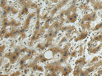
Immunohistochemistry analysis of paraffin embedded Human liver cancer tissue using 216471(DAP3 Antibody) at a dilution of 1/140(Cytoplasm).

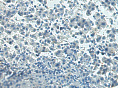
In comparision with the IHC on the left, the same paraffin-embedded Human liver cancer tissue is first treated with the fusion protein and then with 216471(Anti-DAP3 Antibody) at dilution 1/140.

The image on the left is immunohistochemistry of paraffin-embedded Human tonsil tissue using 216471(Anti-DAP3 Antibody) at a dilution of 1/140.

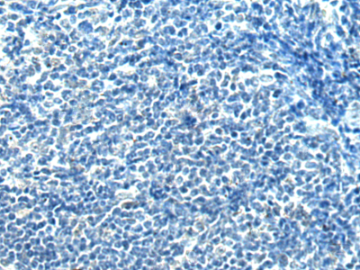
In comparision with the IHC on the left, the same paraffin-embedded Human tonsil tissue is first treated with fusion protein and then with D220543(Anti-DAP3 Antibody) at dilution 1/140.

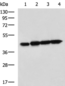
Gel: 8%SDS-PAGE, Lysate: 40 μg;
Lane 1-4: Jurkat, Hela, A549 and HepG2 cell lysates;
Primary antibody: 216471(DAP3 Antibody) at dilution 1/800;
Secondary antibody: HRP-conjugated Goat anti rabbit IgG at 1/5000 dilution;
Exposure time: 1 minute
          Publications