Email: info@neweastbio.com | Call: 6109452007


About NewEast
NewEast Biosciences pioneered the research and development of the antibodies for GTPases and mutated Oncogene ten years ago. GTPases involve (1) signal transduction in response to activation of cell surface receptors, including transmembrane receptors such as those mediating taste, smell and vision, (2) protein biosynthesis at the ribosome, (3) regulation of cell differentiation, proliferation, division and movement, (4) translocation of proteins through membranes, (5) transport of vesicles within the cell, and vesicle-mediated secretion and uptake, through GTPase control of vesicle coat assembly. An oncogene is a gene that has the potential to cause cancer.
We offer three unique categories of antibodies, which (1) recognize only the active configuration of GTPase (not the inactive one), (2) mutated Oncogene (not mild type) and (3) have super affinity for cAMP and cGMP (no acetylation required). We have over one thousand peer reviewed articles cited our products.

Citations on Top 50 Journals
(Google Scholar)
- Rank Journal Name
- 1. Nature
- 7. Nature Communications
- 8. Cell
- 13. Proceedings of the National Academy of Sciences
- 18. Nucleic Acids Research
- 20. Nature Medicine
- 25. ACS Nano
- 29. Scientific Reports
- 33. PLoS ONE
- 34. Science Advances
- 37. Nature Genetics
- 41. International Journal of Molecular Sciences
- 49. Frontiers in Immunology
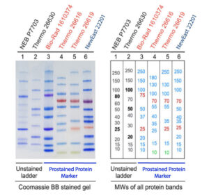
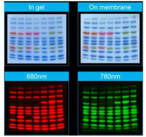 The images are produced from a single controlled experiment. All bands except orange ones are well compatible with near-infrared (NIR) fluorescent system. Note that the red band in Thermo’s product cannot be scanned by NIR fluorescence either. The cat. # of protein markers (left to right) is 22201, 22301, 22202, 22302, 22101, 22203 and 22204.
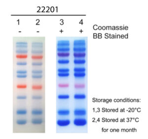
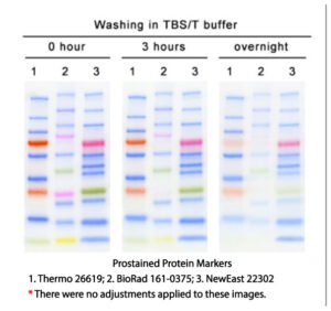 The membrane (PVDF or NC) after transferring during western blot experiment needs to undergo blocking, primary and secondary antibody incubation, and a multi-step washing process in between. It can take as short as 3 hours or even overnight. In a controlled experiment, our marker and ones of Thermo and Biorad are subjected to 3 hours to overnight washing with TBST washing buffer. After 3 hours, all bands of our products are very clear while the brightness of Thermo’s 10 kD band has been extremely weak and Biorad’s 25K band has completely disappeared, and its 10 KD band is also extremely weak. After overnight washing, only 25K and 20K of our marker are weakened, and the rest of the bands are still very clear. In comparison, Thermo’s 10 KD band is almost completely disappeared, and the two red bands is very faint. More than half of the bands for Biorad product disappears or becomes very faint.
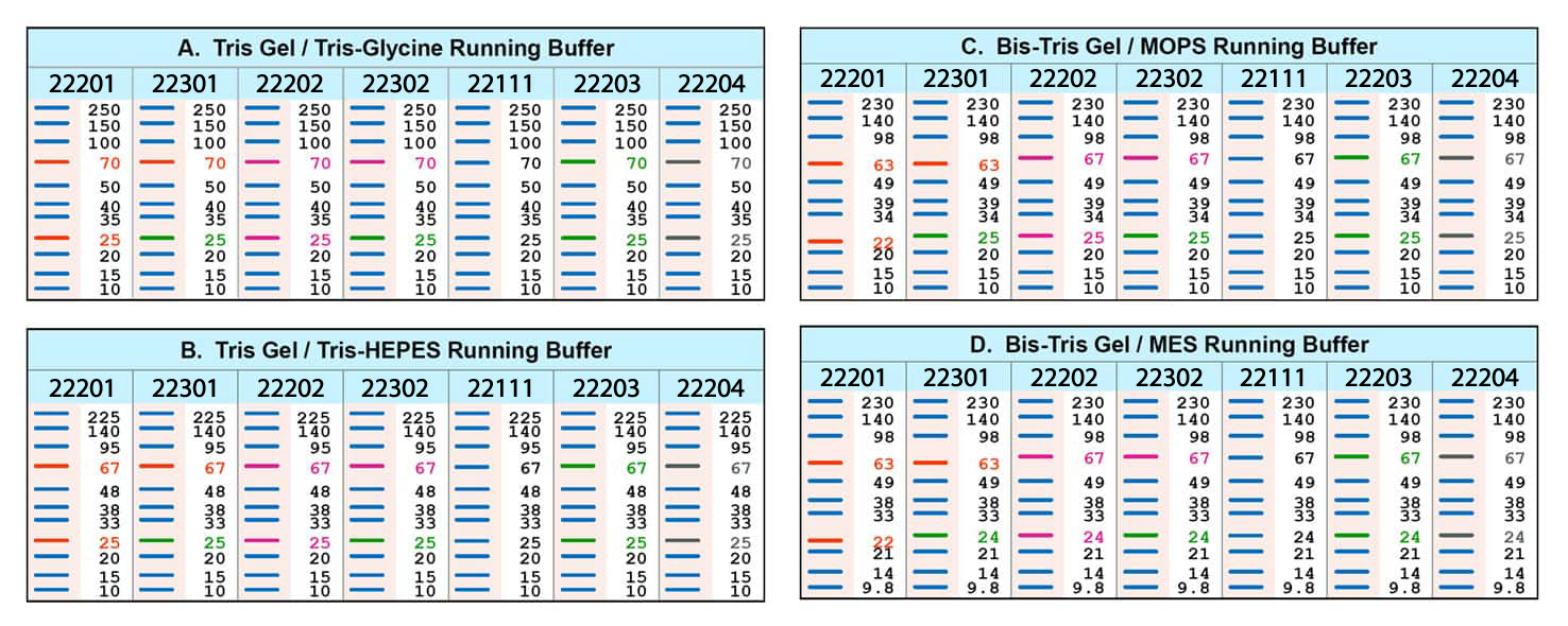 The migration of the prestained marker proteins during electrophoresis depends not only on the proteins themselves but also on the coupled dyes. Therefore, the markers usually show different migration patterns in different electrophoresis systems. The MWs of them are originally calibrated in Tris gel/Tris-Glycine buffer system (Table A). The shifts of the markers in other electrophoresis systems are shown in Table B-D for references. We highly recommend to specifically calibrate their MWs by unstained protein MW standard in corresponding systems other than Tris gel/Tris-Glycine buffer.