| Anti-GαiGTP Mouse Monoclonal Antibody |
| Gene Symbol: Gnai |
| Description: Anti-Gαi-GTP Mouse Monoclonal Antibody |
| Background: Heterotrimeric G proteins are critical cellular signal transducers. Gαi represents one sub-family of G proteins that could mediate the inhibition of adenylyl cyclases. Other biochemical and physiological functions of Gα i proteins are being explored. |
| Immunogen: Recombinant full length protein of active Gα i1 |
| Applications: IP, IHC and IF (Not applicable for WB since SDS denatures Gαi GTPase) |
| Published Applications: IP, IHC and IF – Click for Details |
| Recommended Dilutions:
IP: 1 µg for 1~2 mg total cellular proteins
IHC, IF: 1:50-1:250 |
| Concentration: 1 mg/ml |
| Host Species: Mouse |
| Format: Liquid |
| Clonality: Monoclonal |
| Isotype: IgG1 |
| Purity: Purified from ascites |
| Preservative: No |
| Constituents: PBS (without Mg 2+ and Ca 2+ ), pH 7.4, 150 mM NaCl, 50% glycerol |
| Species Reactivity: Anti-active Gα i antibody recognizes active Gα i1, Gα i2, and Gα i3 of vertebrates. |
| Storage Conditions: Store at -20°C. Avoid repeated freezing and thawing |
Immunoprecipitation/Western blot:
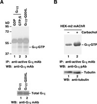 |
Gαi activation assay.
A. CHO cells were transfected with wild type Gα i1 (lanes 1 and 2) or constitutively active Gα i1-Q204L (lane 3). Cell lysates were treated with GDP(lane 1)or GTPγS (lane 3). Lysates were then incubated with an anti-active Gα i monoclonal antibody (Cat. No.26901) (top panel). The precipitated active Gα i was immunoblotted with an anti-Gα i monoclonal antibody (Cat. No. 26003). The bottom panel shows the Western blot with anti-Gα i monoclonal antibody (Cat.No. 26003) of the cell lysates.
B. HEK293 cells stably expressing human m2 mAChR were treated with (lane 2) or without (lane 1)carbachol. Cell lysates were then incubated with an anti-active Gα i monoclonal antibody (Cat. No. 26901) (top panel).The precipitated active Gα i was immunoblotted with an anti-Gα i rabbit polyclonal antibody (Cat. No. 21006). The bottom panel shows the Western blot with anti-tubulin of the cell lysates. |
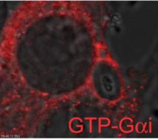 Confocal microcopy images of RAW 264.7 cells expressing YFP-Gαi2QL or YFP-Gαi2 immunostained for GTP-Gαi delineated by phalloidin-Alexa Fluor 647 staining Confocal microcopy images of RAW 264.7 cells expressing YFP-Gαi2QL or YFP-Gαi2 immunostained for GTP-Gαi delineated by phalloidin-Alexa Fluor 647 staining |
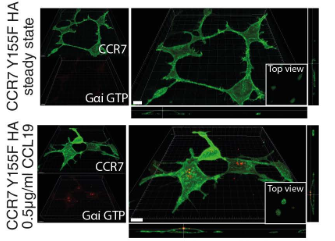 G-protein activation in HEK293 cells expressing CCR7 variants was visualized by confocal microscopy using an antibody specifically recognizing active, GTP-bound Gαi G-protein activation in HEK293 cells expressing CCR7 variants was visualized by confocal microscopy using an antibody specifically recognizing active, GTP-bound Gαi |
|